设为首页 | 加入收藏
当前位置:主页 > 专业资源 > 技术资料 >
技术资料
R&D Technical Information - Bioassay
发布时间:2014-11-13 13:28 来源:未知

Chemotaxis Bioassay

Introduction

R&D Systems uses chemotaxis bioassays to measure the activity of the following chemokines and neutralizing antibodies:

Human Chemokines 
6Ckine Fractalkine MCP-3 MPIF-1
CCL28 HCC-1 MCP-4 MPIF-2
CTACK I-309 MDC PARC
CXCR3 I-TAC MIG RANTES
CXCR4 IP-10 MIP-1 alpha SDF-1 alpha
Eotaxin MCP-1 MIP-1 beta SDF-1 beta
Eotaxin-3 MCP-2 MIP-3 beta  

Mouse Chemokines

6Ckine Eotaxin MARC MIP-3 beta
C10 Fractalkine MCP-5 MPIF-2
CCL28 I-TAC MIG RANTES
CRG-2 JE MIP-1 alpha SDF-1 alpha
CTACK Lymphotactin MIP-1 beta TCA-3

Chemotaxis Bioassay Materials


Cell Preparation
  • Alcohol
  • 2X PBS
  • 1X PBS
  • Percoll (Amersham Pharmacia BioTech Catalog # 17-0891-01)
  • RPMI 1640 (Irvine Catalog # 9160), supplemented 
    2 mM L-glutamine 
    100 U/mL Penicillin 
    100 mg/mL Streptomycin
  • DMEM high glucose (Irvine Catalog # 9024),supplemented 
    2 mM L-glutamine
    100 U/mL Penicillin
    100 mg/mL Streptomycin
  • Fetal Bovine Serum (Sigma Catalog # F2442)
  • G-418 (Gentamycin sulfate) (Sigma Catalog # G1264)
  • 2-mercaptoethanol
  • Ficoll-Hypaque M (1.119 mg/mL final density)
    Ficoll type 400 (Sigma Catalog # F4375)
    Hypaque M-76 (Nycomed Catalog # NDC 0407-0776-04)
    1. Make a 9% Ficoll type 400 solution with ddH20
    2. Combine 900 mL 9% Ficoll + 200 mL Hypaque M-76
    3. Sterilize
    4. Check density
Cell Growth
  • Fresh lymphocytes: purified from fresh blood or leukocyte units - use to test rhFractalkine, rhMPIF-1, rhPARC, rmFractalkine
  • Growth Medium: RPMI 1640, supplemented
    10% Fetal Bovine Serum (Sigma Catalog # F2442)
  • Cultured lymphocytes: purified from fresh blood or leukocyte units and cultured - use to test rh6Ckine, rhCXCR4, rhMIP-3 beta, rhSDF-1 alpha, rhSDF-1 beta, rm6Ckine, rmMIP-3 beta, rmSDF-1 alpha
  • Growth Medium: RPMI 1640, supplemented
    10% Fetal Bovine Serum (Sigma Catalog # F2442)
    50 µM 2-mercaptoethanol
  • IL-2 Cultured lymphocytes: purified from fresh blood or leukocyte units and cultured - use to test rhCXCR3, rhI-TAC, rhIP-10, rhMDC, rhMIG, rmCRG-2, rmI-TAC, rmLymphotactin, rmMIG
  • Growth Medium: RPMI 1640, supplemented
    10% Fetal Bovine Serum (Sigma Catalog # F2442)
    50 µM 2-mercaptoethanol
    10 ng/mL rhIL-2 (R&D Systems Catalog # 202-IL)
  • Monocytes: purified from fresh blood or leukocyte units and cultured 2 days to allow detachment from flask - use to test rhHCC-1, rhMCP-1, rhMCP-2, rhMCP-3, rhMCP-4, rhMIP-1 alpha, rhMIP-1 beta, rhRANTES, rmC10, rmJE, rmMARC, rmMCP-5, rmMIP-1 alpha, rmMIP-1 beta, rmRANTES
  • Growth Medium: RPMI 1640, supplemented
    10% Fetal Bovine Serum (Sigma Catalog # F2442)
  • Y3-hCCR3 Cells: human CCR3 transfected rat myeloid cell line obtained from Dr. Albert Zlotnik at DNAX - use to test rhEotaxin, rhEotaxin-3, rhMPIF-2, rmEotaxin, rmMPIF-2
  • Growth Medium: DMEM, supplemented
    10% Fetal Bovine Serum (Sigma Catalog # F2442)
    25 mM HEPES (GIBCO Catalog # 15630-080)
    1 mg/mL G-418 (Sigma Catalog # G1264)
  • BW5147.G.1.4.OUAR.1 Cells: AKR/J mouse T cell lymphoma cell line, ATCC Catalog # CRL-1588 - use to test rhI-309, rmTCA-3. TIB-48 cells (BW5147.G.1.4 thioguanine resistant ATCC cell line) can also be used
  • Growth Medium: DMEM, supplemented
    10% Fetal Bovine Serum (Sigma Catalog # F2442)
  • mCCR10 Transfected BaF/3 Cells: mouse CCR10 transfected, IL-3 dependent cell line derived from mouse B cells - use to test rhCCL28, rhCTACK, rmCCL28, rmCTACK
  • Growth Medium: RPMI 1640, supplemented
    10% Fetal Bovine Serum (Sigma Catalog # F2442)
    50 µM 2-mercaptoethanol
    10 ng/mL rmIL-3 (R&D Systems Catalog # 403-ML)
    10 mM HEPES (GIBCO Catalog # 15630-080)
    1 mg/mL G-418 (Sigma Catalog # G1264)

Procedure

Cell Growth and Preparation
As with all materials of human source, gloves and lab coats should be worn. All materials contaminated by these cells should be either decontaminated or disposed of in biohazard containers to be autoclaved. All procedures are carried out under sterile conditions.


Purification of Mononuclear Cells from Fresh Heparinized Human Blood
  1. Obtain fresh human heparinized blood.
  2. Using 1 part of Ficoll-Hypaque M, carefully pipette 2 parts of heparinized blood on top of the Ficoll-Hypaque M in a 15 mL centrifuge tube.
    1. Centrifuge 20 minutes at 500 x g, 20 °C. 
      Do not use centrifuge brake.
    2. Carefully remove tubes from centrifuge. There should now be very distinct layering of the blood. The very top layer should be pale yellow and is the plasma portion of blood. The mononuclear cells (MNCs contain lymphocytes and monocytes) are contained in the band just under the plasma which also contains platelets. This band separates the plasma from the ficoll layer. The polymorphonuclear cells (PMNs contain granulocytes, eosinophils and basophils) are suspended in the ficoll-hypaque layer, RBCs make up the lowest layer.
  3. Carefully draw off MNC layer with a plastic pipette, taking some plasma but no PMNs.
  4. Draw off PMNs into another tube. Avoid taking any RBCs (these can be used in the myeloperoxidase assay, for neutrophil chemotaxis or eosinophil chemotaxis).
  5. Wash MNC’s with PBS.
    1. Dilute cells at least 1:4 with 1X PBS and centrifuge 5 minutes at 500 x g.
    2. Repeat this wash until the supernatant is clear and all platelets are removed.
    3. Count cell number and obtain a differential identifying the types and percentage of cells in your prep. There should be <10% granulocytes and no platelets.
  6. Resuspend cells in appropriate Growth Medium to 10 x 106 cells/mL and add to a tissue culture flask. Incubate for one hour at 37 °C. Remove suspended cells (lymphocytes, which are processed further in step 7) and add fresh media to more than cover attached cells in the flask (monocytes). Allow cells to incubate for 48 hours at 37 °C. The monocytes should now be in suspension and are ready to use in the monocyte chemotaxis assay.
  7. Centrifuge lymphocytes. These are now ready to be used in the Fresh Lymphocyte Chemotaxis. For Cultured Lymphocyte Chemotaxis, resuspend to 2 - 3 x 10⁶ cells/mL in appropriate Growth Medium. Incubate for 5 - 8 days (for cultured lymphocytes) or 3 weeks (for IL-2 cultured lymphocytes) prior to using in chemotaxis. Replace media when cells are overcrowded and media spent. The media for the IL-2 cultured lymphocytes will need to be changed every week to replenish nutrients and remove waste.
  8. Larger numbers of lymphocytes/monocytes can be obtained through the purchase of leukocyte units from your community blood center. Cell population separation must then be performed using a pre-formed Percoll gradient and will not occur using the above Ficoll-Hypaque M. Refer to the section below on cell purification from leukocyte units.
  9. Lesser amounts of cells need to be purified if the 48-well microchemotaxis chamber is used. Detection of cell chemotaxis is accomplished by counting the number of cells migrated per well through a microscope (usually this is the average count per 4 - 5 high power fields).
    Purification of Monocytes and Lymphocytes from
Leukocyte Units
  1. Cut the alcohol tube protruding from blood unit with a pair of scissors and allow blood to flow into a sterile 50 mL centrifuge tube.
    Units must be at room temperature.
    Whenever more than one unit of blood is received, process each unit separately.
  2. Centrifuge 20 minutes at 500 x g.
  3. Three distinct layers are formed during centrifugation. The top layer is plasma. The middle layer is white blood cells and the bottom layer consists of red blood cells. Remove and discard most of plasma layer. Transfer WBC layer and top of RBC layer to a 50 mL centrifuge tube and dilute to 50 mL with 1X PBS. Remaining RBC layer can be discarded.
  4. To concentrate the WBC layer, repeat centrifugation procedure 3 or 4 times. This will remove the majority of RBCs. Remove platelets from the preparation by centrifuging for 10 minutes at 1600 rpm. Remove and discard supernatant only. Repeat this procedure 3 or more times until platelets no longer cause the supernatant to be cloudy.
  5. Meanwhile, prepare Percoll/PBS gradient tubes: Sterilize and dry 15 mL polycarbonate Sorvall centrifuge tubes using alcohol. Add to each tube 3.5 mL of 2X PBS and 4 mL of Percoll (Add sequentially in any order. The two solutions may mix during addition, but do not purposely mix). Centrifuge for 40 minutes at 16000 rpm using a Sorvall SS-34 rotor.
  6. Resuspend WBC pellet with 1X PBS (resuspend in only enough volume to add to all tubes made) and carefully overlay 2 mL of suspension onto pre-formed Percoll gradients.
  7. Centrifuge for 20 minutes at 2000 rpm.
  8. Four distinct layers are formed during centrifugation. From top to bottom they are platelets, monocytes, lymphocytes/monocytes, granulocytes/red cells. Carefully remove and discard the platelet layer. Remove and transfer monocyte layer of each Sorvall tube to a labeled 50 mL tube. Remove and transfer the lymphocyte/monocyte layer of each Sorvall tube to another labeled 50 mL tube. Discard granulocytes/red cells.
  9. Dilute both cell preparations with 1X PBS to 50 mL. Remove a 0.5 mL aliquot and count cells to determine cell number and differential.
  10. Remove any remaining platelets from cell preparations by centrifuging suspension for 5 - 10 minutes at 1600 rpm. Repeat procedure 2 or more times.
  11. Based on cell count, resuspend cell pellet to 2 - 4 x 10⁷ cells/mL in appropriate Growth Medium and seed T-175 flasks (1 each for monocyte and lymphocyte/monocyte preparations).
  12. Allow cells (monocytes) to attach to flask. Incubate for 1 hour at 37 °C in a 5% CO2 humidified chamber. Gently pour off suspended lymphocytes into another flask (continue with step 13) and add back appropriate Growth Medium. Return flask to incubator and use cells in monocyte chemotaxis assay 2 days later when monocytes begin growing in suspension.
  13. Centrifuge lymphocytes. These are now ready to be used in the Fresh Lymphocyte Chemotaxis. For Cultured Lymphocyte Chemotaxis, resuspend to 2 - 3 x 10⁶ cells/mL in appropriate Growth Medium. Incubate for 5 - 8 days (for cultured lymphocytes) or 3 weeks (for IL-2 cultured lymphocytes) prior to using in chemotaxis. Replace media when cells are over crowded and media spent. The media for the IL-2 cultured lymphocytes will need to be changed every week to replenish nutrients and remove wastes.


Y3-hCCR3 Cells
Doubling Time: Approximately 16 hours.
Appearance: Cells are primarily attached. As cell density increases, a larger percentage will be suspended in culture.
  1. Seed cells at 5 x 10⁴ cells/mL in appropriate Growth Medium.
  2. Split cells every 3 - 4 days and re-seed in fresh media.
  3. Use cells when they have just become confluent and have had fresh media added within the past 24 - 48 hours.
BW5147.G.1.4 Cells
Doubling Time: Approximately 16 hours.
Appearance: Cells are primarily attached. As cell density increases, a larger percentage will be suspended in culture.
  1. Seed cells at 5 x 10⁴ cells/mL in appropriate media.
  2. Split cells every 3 days and re-seed in fresh media.
  3. Use cells when they have just become confluent and have had fresh media added within the past 24 - 48 hours.

Mouse BaF/3 Cells Transfected with mCCR10
Doubling Time: Approximately 18 hours.
Appearance: Cells appear suspended.
  1. Seed cells at 5 x 10⁴ cells/mL in appropriate Growth Medium.
  2. Split cells every 3 - 4 days and re-seed in fresh Growth Medium.
  3. Use cells when they are in log phase and have had fresh media added within the past 24 hours.

Chemotaxis Assay
Chemotaxis Assay using the standard 96-well Chemotaxis Chamber and using MTT or Alamar Blue to measure the number of cells migrating through the membrane.
  1. Make serial dilutions of samples in Assay Medium.
    1. Add 100 µL Assay Medium to each well of 96-well plate.
    2. Leave column 12 as a blank (cells + media only control).
    3. Add 50 µL of sample or standard to appropriate row of column 1. Serially dilute by mixing the 50 µL 4 - 5 times and transferring 50 µL to next well in row. Repeat to column 11. Remove and discard last 50 µL.
    4. Set up antibodies as per package insert; preincubate serial dilutions of the antibody with cytokine (at a concentration where the final concentration is at maximal chemotaxis). Cytokine only and antibody only control wells are required.
    5. Antibodies to cell surface receptors are set up with constant amount of cytokine in lower chamber at a final concentration to be at maximal chemotaxis and addition of serially diluted antibody to the top chamber with cells
  2. Transfer 75 µL of volume from sample plate to appropriate wells of the 96-well chemotaxis chamber. See manufacturer’s instructions on assembly, care and cleaning of these chambers. A clean chamber is important in obtaining good results.
  3. Assemble chamber as per manufacturer’s instructions using the 5 µm pore size PVP-free polycarbonate filter (8 µm for monocyte chemotaxis). Avoid forming bubbles as they will cause variation in reading the plate.
  4. Resuspend cells to 20 x 106 cells/mL (10 x 106 cells/mL for monocyte and BaF/3 chemotaxis) in Assay Medium. Add 100 µL to upper wells of chamber. Avoid forming bubbles at the level of the filter. Incubate for 3 hours at 37 °C.
  5. Disassemble chamber and remove filter. Scrape cells that did not migrate from the top of the filter. Stain the filter with the Hema 3 Staining System per manufacturer’s instructions. Read O.D. of stained well filters (the 96-well filter will snap into place) per manufacturer’s instructions.
  6. Carefully mix media remaining in lower chemotaxis chamber and transfer to a new 96-well plate.
    1. If using MTT to develop the chemotaxis assay, add 20 µL of MTT (at 5 mg/mL) to transferred cells. Incubate for 4 hours at 37 °C. Add 100 µL of Solubilization Buffer to the wells, incubate for 4 hours and read on a plate reader at 540/690 nm. Graph using 4-parameter fit equation.
    2. If using Alamar Blue, add 10 µL of Alamar Blue (100 µg/mL) to transferred cells. Incubate for at least 4 hours (may incubate overnight) at 37 °C. Read plate on a fluorescent plate reader, excitation at 544 nm and emission at 590 nm. Graph using 4-parameter fit equation.
      Lymphocytes do not adhere to the underside of the filter and will migrate through to the well below. Extreme care must be taken when disassembling the chamber to avoid variations in lower chamber well volumes. Staining of the filter will ensure that cells did not adhere.

Figure 1
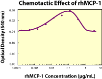
Fig. 1. Human MCP-1 chemoattracts human monocytes, exhibiting a bell-shaped dose response curve. The ED50 for this effect is typically 0.005 - 0.02 µg/mL.
Figure 2
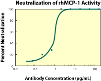
Fig. 2. To measure the ability of the antibody to neutralize the chemoattractant activity of rhMCP-1 for human monocytes, rhMCP-1 was incubated with various concentrations of the antibody for 30 minutes at room temperature in a 96-well microplate. Following this preincubation period, 35 µL of the cytokine-antibody solution (containing rhMCP-1 at a final concentration of 0.1 µg/mL and antibody at the concentrations indicated) was transferred to the lower compartment of a 96-well chemotaxis chamber (NeuroProbe, Cabin John, MD). The chemotaxis chamber was then assembled using a PVP-free polycarbonate filter (8 micron pore size) and 1 x 10⁶ cells/well was added to the top chamber. After incubation for 75 minutes at 37 °C in a 5% CO2 humidified incubator, the chamber was disassembled and the filter was fixed and stained using Leukostat (Fisher Scientific). The optical density of the filter, which is proportional to the number of cells that migrated across the filter, was then read in a microplate reader set at 540 nm. As shown in figure 2, the ND50 for this lot of antibody is approximately 0.8 - 2.5 µg/mL.

Troubleshooting

  1. Using glass will activate platelets and cause the release of factors affecting WBC integrity and release. Using glass will also result in the release of chemokines that will affect chemotaxis.
  2. Ensure that the chemotaxis chamber is clean and dry.
  3. Bubbles in either the lower or upper parts of the chamber will inhibit migration.
  4. Disassemble the chamber carefully so that the volume in the lower chamber remains uniform.
  5. Failure to adequately remove all cells that have not migrated from the top of the filter will cause abnormal staining.

References


T Cell Chemotaxis
1.Bacon, K.B. et al. (1988) Br. J. Pharmacol. 95:966.
2.Falk, W. et al. (1980) J. Immunol. Methods 33:239.
3.Tanaka, Y. et al. (1993) Nature 361:79.
4.Taub, D.D. et al. (1993) J. Exp. Med. 177:1809.
4.Bluel, C.C. et al. (1996) Nature 382:829.
6.Oberlin, E. et al. (1996) Nature 382:833.

Neutrophil Chemotaxis
7. Magazin, M. et al. (1992) Eur. Cytokine Netw. 3:461. 

Eosinophil Chemotaxis 
8. Rothenberg, M.E. et al. (1995) Proc. Natl. Acad. Sci. USA 92:8960.
9. Dahinden, C.A. et al. (1994) J. Exp. Med. 179:751.
10. Weber, M. et al. (1995) J. Immunol. 154:4166.
11. Rot, A. et al. (1992) J. Exp. Med. 176:1489.
12. Garcia-Zepeda, E.A. et al. (1996) Nature Med. 2:449. 

Myeloid Progenitor Chemotaxis
13. Patel, V. et al. (1997) J. Exp. Med. 185:1163.

Monocyte Chemotaxis 
14. Matsushima, K. et al. (1989) J. Exp. Med. 169:1485.
15. Rollins, B. et al. (1989) Blood 78:111


Proliferation Bioassay HUVECs

Introduction

R&D Systems uses HUVECs (Human Umbilical Vein Endothelial Cells) in proliferation bioassays of the following cytokines and neutralizing antibodies:

Human Cytokines

  • PD-ECGF
  • VEGF R1 (Flt-1)/Fc chimera
  • VEGF121
  • VEGF R2 (KDR)/Fc chimera
  • VEGF165
  • VEGF/PlGF

Mouse Cytokines

  • VEGF120
  • VEGF R1 (Flt-1)/Fc chimera
  • VEGF164
  • VEGF R2 (Flk-1)/Fc chimera

Rat Cytokines

  • VEGF164

HUVEC Bioassay Materials

Cell Growth and Preparation

  • HUVEC primary culture (Clonetics Catalog # CC-2519)
  • Growth Medium: Endothelial Cell Growth Medium (Clonetics Catalog # CC-3024)
    2% Fetal Bovine Serum (JRH Catalog # 12103-78P)
Plate Preparation


  • 96-well flat bottom microtiter plate
  • Dulbecco's PBS (Irvine Catalog # 9240)
  • Collagen Solution: 40 µg/mL Type I Collagen (Sigma Catalog # C7661)
    0.15 M Acetic Acid
    Leave overnight at room temperature to dissolve
    Add 1/25 volume of 3.76% NaCl
    Store solution at 4 °C
HUVEC Bioassay
  • HUVECs
  • 3H-thymidine (NEN Catalog # NET027E)
  • Dulbecco's PBS (Irvine Catalog # 9240)
  • Bovine Serum Albumin (Sigma Catalog # A-7888)
    1X Trypsin Solution: Dilute 10X Trypsin EDTA (Irvine Catalog # 9342) 1:10 in PBS
  • Assay Medium: Medium-199 1X Earle's Salts (Invitrogen # 11150-059)
    10% Fetal Bovine Serum (JRH Catalog # 12103-78P), heat inactivated 
    10 mM HEPES
    100 units/mL Penicillin 100 µg/mL Streptomycin

Procedure

Cell Growth and Preparation
As with all materials of human source, gloves and lab coats should be worn. All materials contaminated by these cells should be either decontaminated or disposed of in biohazard containers to be autoclaved. All procedures are carried out under sterile conditions.

Doubling Time: Approximately 30 hours
Appearance: Attached


  1. Seed cells at 3 x 10⁵ cells/75 cm2 flask.
  2. Feed cells every 3-4 days with fresh Growth Medium.
  3. Split cells every week and reseed in fresh Growth Medium.
    Cultures are quality assured for up to seven passages.
Plate Preparation
  1. Add 50 µL/well of Collagen Solution.
  2. Incubate for 2 hours at room temperature. Alternatively, incubate at 4 °C overnight.
  3. Wash plate twice with PBS. Coated plates may be decanted, air dried and stored at -20 °C for up to 30 days.

HUVEC Bioassay

  1. Reconstitute standards and samples in PBS + 1.0 mg/mL BSA.
  2. Dilute standards and samples to working concentration with Assay Medium.
  3. Add 50 µL of Assay Medium to each well of a 96-well plate.
  4. Add standards and samples to the plate. Add 25 µL to first well and serial dilute. Leave last well with dilution media only, as a blank. Run samples in duplicate. Refer to figure 2 legend for neutralizing antibody assay conditions.
  5. Add cells to all wells. Harvest cells by trypsinization with 1X Trypsin Solution. Wash cells twice with Medium-199. Resuspend cells in Assay Medium at 1 x 10⁵ cells/mL. Add 50 µL of cells to each well.
  6. Incubate for 72 hours at 37 °C with 5% CO2 in a humidified chamber.
  7. Pulse last 18-24 hours with 0.5 µCi 3H-thymidine. Prepare 50.0 µCi/mL 3H-thymidine working stock in Assay Medium. Add 10 µL/well of working stock.
  8. Harvest cells and count. Wash plate with 100 µL/well PBS. Add 100 µL/well of 1X Trypsin Solution to detach cells. Harvest on glass fiber filters using cell harvester. Count 3H-thymidine incorporated into DNA. Plot the dose response using a 4-parameter fit equation.

Troubleshooting

  1. Use lower concentration of trypsin and treat for approximately 2 minutes. Do not over trypsinize.
  2. Coat plate with collagen prior to assay.
  3. Only use cells up to passage seven.
  4. Keep timing of assay consistent.
  5. Always include the correct controls.

References

  1. Conn, G. et al. (1990) Proc. Natl. Acad. Sci. USA 87:1323.
  2. Usuki, K. et al. (1990) Cell Regul. 1:577.
Figure 1. Human VEGF stimulates the 3H-thymidine incorporation by human umbilical vein endothelial cells in a dose-dependent manner. The ED50 for this effect is typically 2-6 ng/mL.
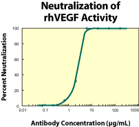
Figure 2. To measure the ability of the antibody to neutralize the bioactivity of rhVEGF on human umbilical vein endothelial cells, rhVEGF was incubated with various concentrations of the antibody for 1 hour at 37 °C in a 96-well microplate. Following this preincubation period, HUVECs were added. The assay mixture in a total volume of 100 µL, containing antibody at the concentrations indicated, rhVEGF at 10 ng/mL and cells at 0.5 x 10⁴ cells/well, was incubated at 37 °C for 72 hours in a humidified CO2 incubator. 3H-thymidine was added during the final 24 hours of incubation. The cells were subsequently harvested onto glass fiber filters and the 3H-thymidine incorporated into DNA was determined. The ND50 of the antibody is approximately 3-6 µg/mL.

Proliferation Bioassay NR6R 3T3 Cells

First Published in R&D Systems 2003 Catalog

Introduction

R&D Systems uses NR6R-3T3 cells in proliferation bioassays to measure the activity of the following cytokines and neutralizing antibodies:

Human Cytokines

  • ß-ECGF
  • FGF acidic
  • FGF basic
  • FGF-4
  • FGF-5
  • FGF-6
  • FGF-8b
  • FGF-8c
  • FGF-17
  • FGF-18
  • PDGF
  • PDGF-AA
  • PDGF-BB
  • PDGF-AB

Bovine Cytokines

  • FGF acidic
  • FGF basic
  • PDGF
  • PDGF-BB

NR6R-3T3 Cell Bioassay Materials

Cell Growth and Preparation
  • NR6R-3T3 cells, a mouse fibroblast cell line (Dr. Angie Rizzino1)
  • 1X Trypsin Solution: Dilute 10X Trypsin EDTA (Irvine Catalog # 9342) 1:10 in PBS
  • Growth Medium: DMEM high Glucose (Irvine Catalog # 9024) 10% Fetal Bovine Serum (Biocell Catalog # 6201)
    2 mM L-glutamine
    100 U/mL Penicillin
    100 µg/mL Streptomycin
NR6R-3T3 Cell Bioassay
  • NR6R-3T3 cells
  • DMEM high Glucose (Irvine Catalog # 9024)
  • Fetal Bovine Serum (Biocel Catalog # 6201)
  • Defibrinated bovine plasma (Irvine Catalog # 1067)
  • PBS (GIBCO Catalog # 14040-133)
  • PBS containing 1 mg/mL BSA (Bayen/Pentex Catalog # 18-068-3)
  • Dialysis tubing (Baxter Catalog # D1614-13, 12,000-14,000 MW cut off)
  • Sodium Heparin (Sigma Catalog # H3149)
  • 3H-thymidine (NEN Catalog # NET027E)
  • Plasma derived bovine serum (PDS) - dialyzed to remove growth factors:
    1. Thaw approximately 100 mL of defibrinated bovine plasma.
    2. Prepare dialysis tubing by cutting it to the appropriate length and softening it in deionized water.
    3. Fill dialysis tubing with defibrinated bovine plasma and seal.
    4. Place dialysis tubing into DMEM. Stir gently on a stir plate for approximately 6 hours at 4 °C. Change the medium by replacing with fresh medium. Dialyze overnight at 4 °C.
    5. Collect dialyzed PDS after 24 hours and filter 2 times through a 0.2 µm filter.
    6. Store extra PDS at -20 to -80 °C.
  • Sodium Heparin Stock
    1. Dissolve sodium heparin in PBS.
    2. Add the sodium heparin solution to DMEM containing 2% PDS (see specific cytokine insert for required final concentration).
  • Growth Medium: DMEM high Glucose 10% Fetal Bovine Serum
    2 mM L-glutamine
    100 U/mL Penicillin
    100 µg/mL Streptomycin
  • Assay Medium: DMEM high Glucose 2% PDS 
    2 mM L-glutamine
    100 U/mL Penicillin
    100 µg/mL Streptomycin
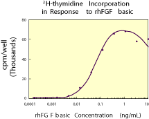
Figure 1. Human FGF basic stimulates the 3H-thymidine incorporation by NR6R-3T3 fibroblasts in a dose-dependent manner.1 The ED50 for this effect is typically 0.1 - 0.25 ng/mL.
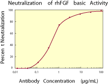
Figure 2. To measure the ability of the antibody to neutralize the bioactivity of rhFGF basic on NR6R-3T3 fibroblasts, human FGF basic was incubated with various concentrations of the antibody for 1 hour at 37 °C in a 96-well microtiter plate. Following this preincubation period, the antigen-antibody mixture was added to quiescent confluent cultures of NR6R-3T3 cells in DMEM with 2% bovine plasma-derived serum. The assay mixture in a total volume of 100 µL, containing antibody at the concentrations indicated and rhFGF basic at 0.5 ng/mL, was incubated at 37 °C for 20 hours in a humidified CO2 incubator. 3H-thymidine was added during the final 2 hours of incubation. The cells were subsequently detached and harvested onto glass fiber filters and the 3H-thymidine incorporated into DNA was determined. The ND50 of the antibody is approximately 0.5 - 1.0 µg/mL.

Procedure

Cell Growth and Preparation
As with all materials of human source, gloves and lab coats should be worn. All materials contaminated by these cells should be either decontaminated or disposed of in biohazard containers to be autoclaved. All procedures are carried out under sterile conditions.

Doubling Time: Approximately 17 hours Appearance: Rectangular shaped, attached cells

  1. Seed 4 x 10⁵ cells into a 75 cm2 flask containing 20 mL Growth Medium.
  2. Feed the cells every 3 days by replacing the medium with fresh Growth Medium.
  3. Split the cells every week and reseed in fresh Growth Medium. The cells are ready to use in the bioassay when they reach confluency (3 x 10⁶ cells/75 cm2 flask).

NR6R-3T3 Cell Bioassay

  1. Add the cells to each well, leaving the last column empty.
    • Harvest the confluent NR6R-3T3 cells by trypsinization with 1X Trypsin solution.
    • Wash the cells twice with 10 mL Growth Medium.
    • Resuspend the cells in Growth Medium at 0.5 x 10⁵ cells/mL.
    • Add 100 µL of cells to each well, leaving the last column empty.
    • Incubate for 2 days (cells should be 90-100% confluent).
    • Shake out the Growth Medium.
    • Gently add 100 µL Assay Medium to each well.
    • Incubate for 24 hours (cells are now in a resting phase and ready to bioassay).
  2. Reconstitute the cytokine (standard) as directed on each specific cytokine insert. If using rhFGF-5, reconstitute samples in PBS containing 1 mg/mL BSA and 0.1 µg/mL sodium heparin. For assays using rhFGF acidic, rhb-ECGF, rhFGF-5 and rhFGF-6, FGF-8b, FGF-8c, FGF-17, and FGF-18 add 10 µL of Sodium Heparin Stock to each well so that the final concentration of sodium heparin in the assay mixture is as follows:
    • rhFGF acidic, rhb-ECGF, and rhFGF-17 – 10 µg/mL sodium heparin
    • rhFGF-5, rhFGF-8b, rhFGF-8c, and rhFGF-18 – 0.1 µg/mL sodium heparin
    • rhFGF-6 – 1.0 µg/mL sodium heparin
  3. Dilute the standards and samples to their working concentration with Assay Medium.
  4. Add the standards and samples to the plate.
    • Add 50 µL to the first well and serial dilute.
    • Leave the last well with Assay Medium only, as a blank control.
Refer to Figure 2 legend for the neutralizing antibody assay conditions for anti-FGF basic antibody (Catalog # AB-233-NA).
  1. Incubate for 16 - 18 hours at 37 °C with 5% CO2 in a humidified chamber.
  2. Pulse the cells with 0.25 µCi 3H-thymidine/well. Incubate for 2 hours.
    • Prepare 25 µCi/mL 3H-thymidine working stock in Assay Medium.
    • Add 10 µL of 25 µCi/mL 3H-thymidine working stock to each well.
  3. Harvest the cells and count.
    • Wash the cells with 100 µL PBS/well.
    • Add 100 µL 1X Trypsin Solution/well to detach the cells.
    • Harvest on glass fiber filters using a cell harvester.
    • While harvesting, incubate the plate in water for 2 minutes to lyse the cells.
    • Wash the plate thoroughly to ensure all the attached cells have been removed.
    • Count the 3H-thymidine incorporated into the DNA.
    • Plot the dose response using a 4-parameter fit equation.

Troubleshooting

  1. Timing is critical for this assay. Longer or shorter incubation times will shift the ED50.
  2. If the cell doubling time changes, it will change the assay results and ED50.

References

  1. Rizzino, A. et al. (1988) Cancer Research 48:4266-4271.


Proliferation Bioassay of TF-1 Cells

Contents

Introduction

TF-1 cells are a factor-dependent human erythroleukemic cell line. TF-1 cells are employed in proliferation bioassays by R&D Systems for the routine quality assurance and quality control of the following cytokines and neutralizing antibodies:

Human Cytokines

  • CNTF
  • Epo
  • GM-CSF
  • IL-3
  • IL-4
  • IL-5
  • IL-13
  • LIF
  • b-NGF
  • SCF
  • OSM

Mouse Cytokines

  • IL-5
  • IL-13
  • SCF

Rat Cytokines

  • CNTF

TF-1 cells will also proliferate in response to human IL-6 and IL-11. TF-1 cells are not responsive to human G-CSF, IL-2, IL-7, IL-9 and M-CSF. The TF-1 cells used by R&D Systems were obtained from Dr. Toshio Kitamura in 1989. Dr. Kitamura has deposited TF-1 cells with ATCC (#CRL-2003). The TF-1 cells deposited by Dr. Kitamura with the ATCC are described as non-responsive to IL-5. If you need to obtain TF-1 cells that will respond to IL-5, please contact R&D Systems Technical Service at 1-800-343-7475.

Procedure

Cell Growth and Preparation

  • TF-1 human premyeloid cell line
  • Growth Medium:
    • RPMI 1640 (Irvine Catalog #9160)
    • 10% (v/v) Fetal Bovine Serum
    • 2 mM L-glutamine
    • 100 units/mL Penicillin
    • 100 µg/mL Streptomycin 2 ng/mL rhGM-CSF (R&D Systems Catalog # 215-GM)

TF-1 Bioassay

  • TF-1 human erythroleukemia cell line
  • 3H-thymidine (NEN Catalog # NET027E)
  • Dulbecco's PBS (Irvine Catalog #9240)
  • BSA (Bayer Pentax Catalog # 81-068-3)
  • Assay Medium:
    • RPMI 1640 (Irvine Catalog # 9160)
    • 10% (v/v) Fetal Bovine Serum
    • 2 mM L-glutamine
    • 100 units/mL Penicillin
    • 100 µ/mL Streptomycin

Cell Growth and Preparation

As with all materials of human source, gloves and lab coat should be worn. All materials contaminated by these cells should be either decontaminated or disposed of in biohazard containers to be autoclaved. All procedures are carried out under sterile conditions.

Doubling Time: Approximately 30 hours.

Appearance: Single cells, slightly irregular in size and shape. A small percentage will attach to the flask.

  1. Seed cells at 5x10⁴ cells/mL or higher in Growth Medium.
  2. Split cells every 3-4 days and reseed in fresh Growth Medium.

TF-1 Bioassay

  1. Reconstitute standards and samples in PBS + 1.0 mg/mL BSA.
  2. Dilute standards and samples to working concentration (6X higher than desired final concentration) with Assay Medium.
  3. Add 50 µL of Assay Medium to each well of 96-well plate.
  4. Add standards and samples to plate.
    • Leave last well with dilution media only as a blank.
    • Run samples in duplicate.
    Note: Refer to figure 2 legend for neutralizing antibody assay conditions.
  5. Add cells to all wells.
    • Resuspend the cells to 2 x 10⁵ cells/mL in Assay Medium.
    • Add 50 µL of cells to each well.
  6. Incubate for 48 or 72 hours at 37°C with 5% CO2 in a humidified chamber.
  7. Pulse 4 hours with 0.5 µCi 3H-thymidine.
    • Prepare 50.0 µCi/mL 3H-thymidine working stock in Assay Medium.
    • Add 10 µL/well of working stock.
  8. Harvest cells and count.
    • Count 3H-thymidine labelled DNA.
    • Plot the dose response using a 4-parameter fit equation.


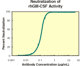
Figure 1. Human GM-CSF stimulates the 3H-thymidine incorporation by TF-1 cells in a dose-dependent manner (Kitamura, T. et al., (1989) J. Cell Physiol. 140(2):323). The ED50 for this effect is typically 0.02 - 0.08 ng/mL. Figure 2. Typical data for anti-hGM-CSF (Catalog # AF-215-NA) is shown. To measure the ability of the antibody to neutralize the bioactivity of rhGM-CSF on human TF-1 cells, rhGM-CSF was incubated with various concentrations of the antibody for 1 hour at 37° C in a 96-well plate. Following this preincubation period, TF-1 cells were added. The assay mixture in a total volume of 100 µL, containing antibody at the concentrations indicated, rhGM-CSF at 0.5 ng/mL and cells at 1 x 10⁵cells/mL, was incubated at 37° C for 48 hours in a humidified CO2 incubator.3H-thymidine was added during the final 4 hours of incubation. The cells were subsequently harvested onto glass fiber filters and the 3H-thymidine incorporated into DNA was determined. The ND50 of the antibody is approximately 0.08 - 0.16 µg/mL.

Troubleshooting

  1. Variations in cell feeding and splitting (growth times) can influence stability of the cell line. Splitting cells at regular intervals and avoiding overgrowth of cells will reduce the possibility of a population of cells becoming GM-CSF growth independent and overtaking the dependent cell population.
  2. Keep timing of assay consistent.
  3. Always include the correct controls.

Reference

  1. Kitamura, T. et al. (1989) Establishment and characterization of a unique human cell line that proliferates dependently on GM-CSF, IL-3 or erythropoietin. J. Cell Physiol.140:323-34.